
Sunday, 4 September 2011
Monday, 6 June 2011
To pronk or not to pronk! The gait of domestic animals.
Pronking is an amusing type of gait that involves leaping into the air with all four feet off the ground. Gazelles are well known to do it when being pursued by a predator and sometimes during play.
Amongst domestic animals lambs and kids (but not adult sheep and goats) can be seen doing it during play. Horses (particularly foals) are seen doing it during play or just to burn up excess energy.
The common types of gaits in seen in our four-legged domestic animals include: walk, trot, pace, canter, and gallop.
At the walk, quadrupeds have one foot raised and the other three feet on the ground, except briefly as they transfer weight from one foot to another. The walk is a four-beat gait. You can clearly hear this as a horse walks along a hard surface.
The trot is a two-beat gait. Diagonally opposite limbs alternate in supporting weight. At a slow trot there are always two diagonally opposite feet on the ground. But at a faster trot there is a phase in the gait when all four feet are off the ground between transferring weight from one pair of limbs to the other.
The Pace is a two-beat gait where the two legs on the same side of the animal move forward together. Compare this to the trot where two diagonally opposite legs move forward together. In both the pace and the trot, two feet are always off the ground. The trot is a much more common gait.
The canter is an asymmetrical, three-beat gait. The sequence of limb impact on the ground is 1-2-1: a hindlimb, then 2 diagonals, followed by a forelimb. The animals can be seen to lead with the left or the right. The leading forelimb is the one that is not part of the diagonal. At faster speeds there will be a suspension phase when all four limbs are off the ground.
The gallop is very much like the canter, except that it is faster. The three-beat canter changes to a four-beat gait. Generally one hind limb hits the ground then the other then one of the front then the other. It is the fastest gait.
Amongst domestic animals lambs and kids (but not adult sheep and goats) can be seen doing it during play. Horses (particularly foals) are seen doing it during play or just to burn up excess energy.
The common types of gaits in seen in our four-legged domestic animals include: walk, trot, pace, canter, and gallop.
 |
| Walking gait |
The trot is a two-beat gait. Diagonally opposite limbs alternate in supporting weight. At a slow trot there are always two diagonally opposite feet on the ground. But at a faster trot there is a phase in the gait when all four feet are off the ground between transferring weight from one pair of limbs to the other.
 |
| Pacing gait |
The canter is an asymmetrical, three-beat gait. The sequence of limb impact on the ground is 1-2-1: a hindlimb, then 2 diagonals, followed by a forelimb. The animals can be seen to lead with the left or the right. The leading forelimb is the one that is not part of the diagonal. At faster speeds there will be a suspension phase when all four limbs are off the ground.
 |
| Galloping gait |
Tuesday, 10 May 2011
Colour and camouflage
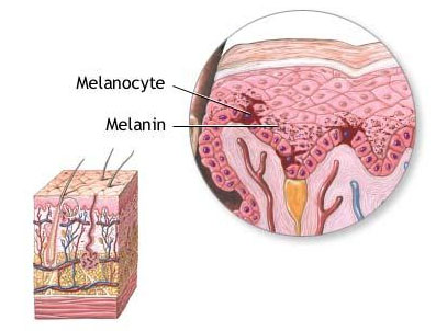 |
| Source: http://training.seer.cancer.gov/melanoma/anatomy/layers.html |
In animals melanin pigments are derived from the amino acid tyrosine. The most common form of biological melanin is a brown-black colour.
However, not all animal colours come from pigments. Structural colours are caused by interference effects rather than by pigments. Colour effects are produced when a surface has microstructures that interfere with the wavelength of light to produce colours. This is analogous to the rainbow colour seen in an oil slick on a wet road and in soap bubbles. This iridescent effect is seen in animals in peacock feathers, butterfly wings and beetle shells.
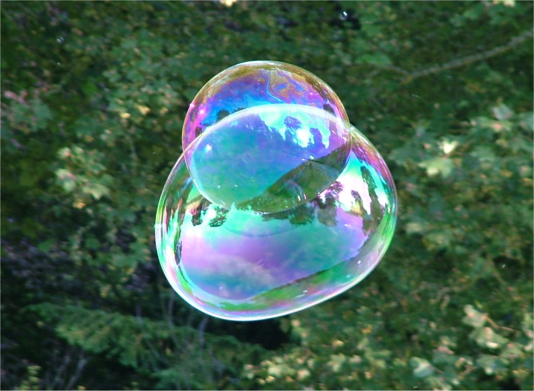 |
Soap bubbles |
 |
| Iridescent peacock feather |
One of the purposes of all this colour is camouflage. Animals use camouflage to blend into their environment and avoid detection. Prey species do this to avoid detection or to confuse their predators. Predator species do it to sneak up on their prey and avoid detection until it is too late.
There are various tactics to achieve camouflage.
Some colour themselves like their background. The brown colours of deer and kangaroo are to match trees or dirt.
 | |
| Euro |
 |
| Ibexes in the Israeli desert |
Some use countershading, where the shading is darker on the back and lighter on the belly. For example, the combination of blue skin and white underbelly of sharks. This makes them difficult to detect from both above and below. From below their lighter belly matches the lighter sea surface. And from above their darker dorsum matches the colour of the deeper water and sea floor.
 |
| White shark |
 |
| Tigress Indrani in Kanha National Tiger reserve of central India. |
More complex patterns can be seen in animals such as flounder, moths, and frogs, among many others. Flounder have a granular pattern which mimics the gravelly surface the rest upon.
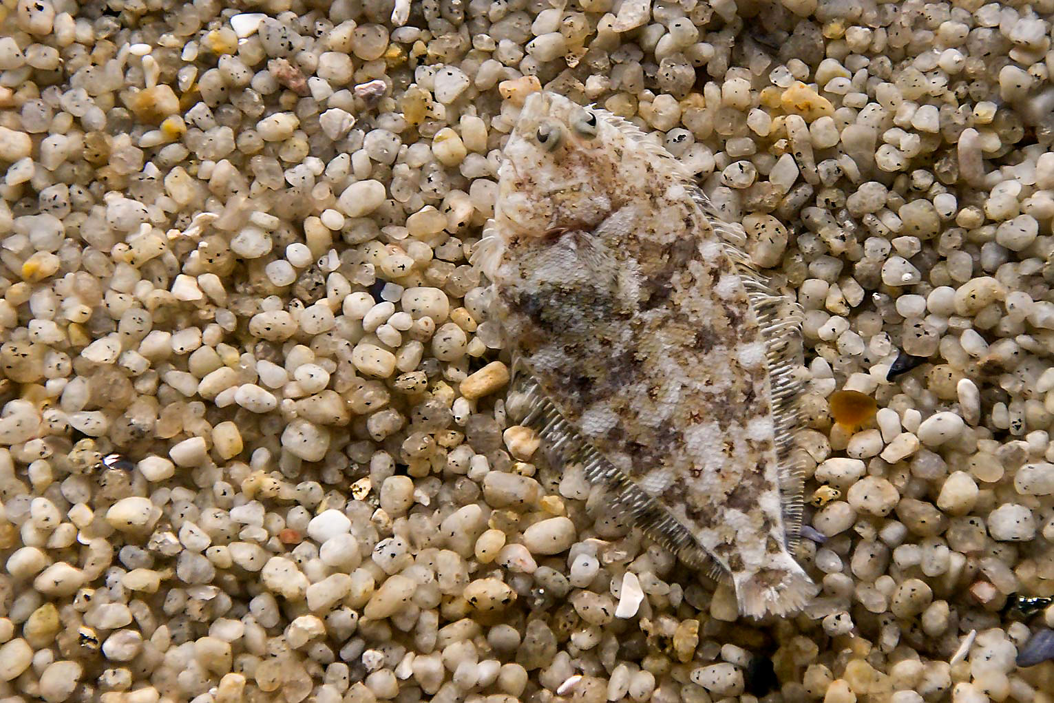 |
| Flounder |
Colouration can be used to break up the outline of an animal's body. For example, in common domestic tabby cats.
 |
| Tabby cat |
 |
| Cuttlefish has changed the pattern on its dorsum to match the gravel it lies upon |
 |
| Stoat colour in summer and winter |
 |
| Leaf insect (Phyllium giganteum) |

Some use patterns or stripes that bled together when animals are in groups. A herd of zebras looks like one large mass, making it difficult for a lion to pick out any individual zebra. This same concept is used by many striped fish species as well.
 |
| Zebras on the Serengeti |
Saturday, 30 April 2011
How vision works

The nerve impulses travel along the optic nerve to the visual cortex area of the brain. The visual cortex area of the brain processes the billions of pieces of information that arrive and form them into a meaningful image for the animal.
Retinal disease
Progressive retinal atrophy (PRA) is a group of genetic diseases seen in certain breeds of dogs and, more rarely, cats. It is characterised by the bilateral degeneration of the retina.
Generalised PRA is the most common form and can be divided into either dysplastic disease, where the cells develop abnormally, and degenerative, where the cells develop normally but then undergo a damaging change. PRA can be further divided into affecting either rod or cone cells.
It causes causes progressive vision loss culminating in blindness. The condition in nearly all breeds is inherited as an autosomal recessive trait.
Friday, 15 April 2011
Why do abscesses smell so bad?

Abscesses may be caused by many different bacteria, fungi, protozoans or by other foreign materials (e.g. a grass seed).
An abscess is a collection of pus (dead neutrophils) that forms a cavity in tissue. It is a defensive reaction designed to prevent the spread of infectious materials. The presence of these organisms damage and kill surrounding cells. The cells release cytokines - chemicals that trigger the inflammatory response. They attract neutrophils and other white blood cells to the area. They also increase the local blood flow resulting in a red appearance (erythema) and feeling of heat. As pus collects, a wall or capsule is formed by the nearby healthy cells in order to prevent the infection spreading to surrounding tissue. This capsule also tends to keep other immune cells from getting at the causative organisms.
This multiplication of organisms and the cellular destruction also forms a cocktail of some unpleasant chemicals. The exact mix depends on the organisms. Gasses such as ammonia, methane and sulphur dioxide (think rotten eggs) may be produced. Two other compounds are largely responsible for the foul odour of putrefying flesh. The delightfully named putrescine and cadaverine, are produced by the breakdown of amino acids in the tissue.
But what is the point of all this stink? To us the smell is disgusting and we are repelled by it. We instinctively avoid anything that creates such odours helping to protect us from exposure to infectious agents.
Thursday, 7 April 2011
Why do nerve impulses only travel in one direction?
 |
| GFP expressing pyramydal cell in mouse cortex. Wei-Chung Allen Lee, et al. |
The fluid inside and outside a cell contains many different kinds of charged particles called ions. Some are negatively charged and some positively. Two important ones are Na+ (sodium) and K+ (potassium).
The resting state
When the neurone is in a resting state it is actually working quite hard. The cell membrane has a structure call a sodium-potassium pump which pumps Na+ to the outside and K+ to the inside of neurones. Because Na+ cannot readily diffuse across the cell membrane a higher concentration of Na+ builds up on the outside of the cell. This redistribution of ion together with the ones from other ions and proteins results in a charge being formed across the membrane - the inside is more negatively charged.

Firing off
When a neurone is stimulated - from an adjacent neurone or an external stimulus (heat, touch) - changes take place in the membrane. Sodium channels in the membrane open briefly and Na+ floods into the cell interior. The inside of the cell changes from negatively charged to positive. This process is known as depolarisation.

Recovering
Just as the sodium channels snap shut potassium channels open and K+ diffuses out of the cell. This causes the charge the inside the cell to head back towards negative again. The sodium-potassium pump works hard and restores the Na+ and K+ to their original sides of the membrane in resting state. This process is known as repolarisation.
This electrical event is called an action potential.

The refractory period
So the cell membrane goes through a cycle of depolarisation and repolarisation. If another stimulus arrives at the membrane during this cycle, the area cannot depolarise again. The current cycle has to finish before it is capable of depolarising again. The refractory period refers to this time when the membrane is not susceptible to depolarising.
And here's why it travels in one direction
An action potential starts at on end of a neurone and spreads as a wave along the it. The currents flowing inwards at one point on the neurone also depolarises the adjacent sections of the membrane. Once an action potential has occurred at a patch of membrane, the membrane patch needs time to recover before it can fire again (the refectory period). For this reason, only the unfired part of the membrane can respond with an action potential. The part that has just fired is unresponsive until the action potential is safely out of range.
Typically the action potential starts at the axon end (by stimulation from another nerve) and travel along a neurone to the synapse end.
Clinical implications
Local anaesthetics are membrane stabilising drugs;. They decrease the rate of depolarisation and depolarisation of neurones. These drugs act mainly by inhibiting sodium influx in the neuronal cell membrane. When the influx of sodium is interrupted, an action potential cannot arise and signal conduction is inhibited.
Disturbances of sodium or potassium levels in the body can directly affect these chemical processes and will interfere with normal nerve conduction.
Low serum potassium levels will often cause a generalised weakness. Increased potassium levels interferes with depolarisation of cells resulting in inability of nerves to fire.
High levels of sodium will cause weakness initially and then with more severe elevations of the sodium level, seizures and coma may occur.
Tuesday, 5 April 2011
What makes eye colour?

In mammals, there are many variations in eye colour: blue, brown, grey, green, and others.
Avian eye colours range from dark brown and yellow through red, blue, and green to metallic silver and gold. In some species, eye colour differs between the sexes.
The superficial (stromal) layers are richly supplied with blood vessels and contain scattered cells with pigment granules. The deepest (epithelial) layer of the iris contains large amounts of dark pigment (melanin).
Blue eyes have the usual deep epithelial layer full of dark pigment but the upper layers are thin and have little or no pigment granules. White light is made up a spectrum of colours. The red wavelengths penetrate and are absorbed in the epithelial layer. The blue wavelengths are scattered by the sparse pigment granules and the iris appears blue.
Brown eyes have lots of pigment granules in the superficial layers and the pigment cells are closely packed. This absorbs most light and the eye appears brown.
Hazel, green and grey irises have progressively less stromal pigment and so show colours in between blue and brown.
The brightly coloured eyes of many bird species are largely determined by other pigments, such as pteridines, purines, and carotenoids.
Albino animals are unable to make the dark pigment melanin. The iris colour is pink as the rich stromal blood supply provides the colour.
Heterochromia is a condition in which one iris is a different colour from the other iris. It usually results due to uneven melanin content and may be inherited or acquired by disease or injury.
 |
| Heterochromia |
 |
| Albino iris |
Subscribe to:
Posts (Atom)